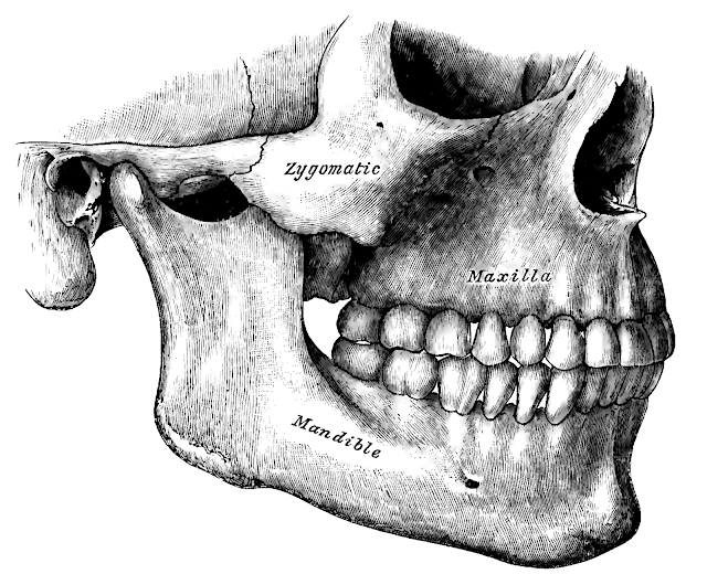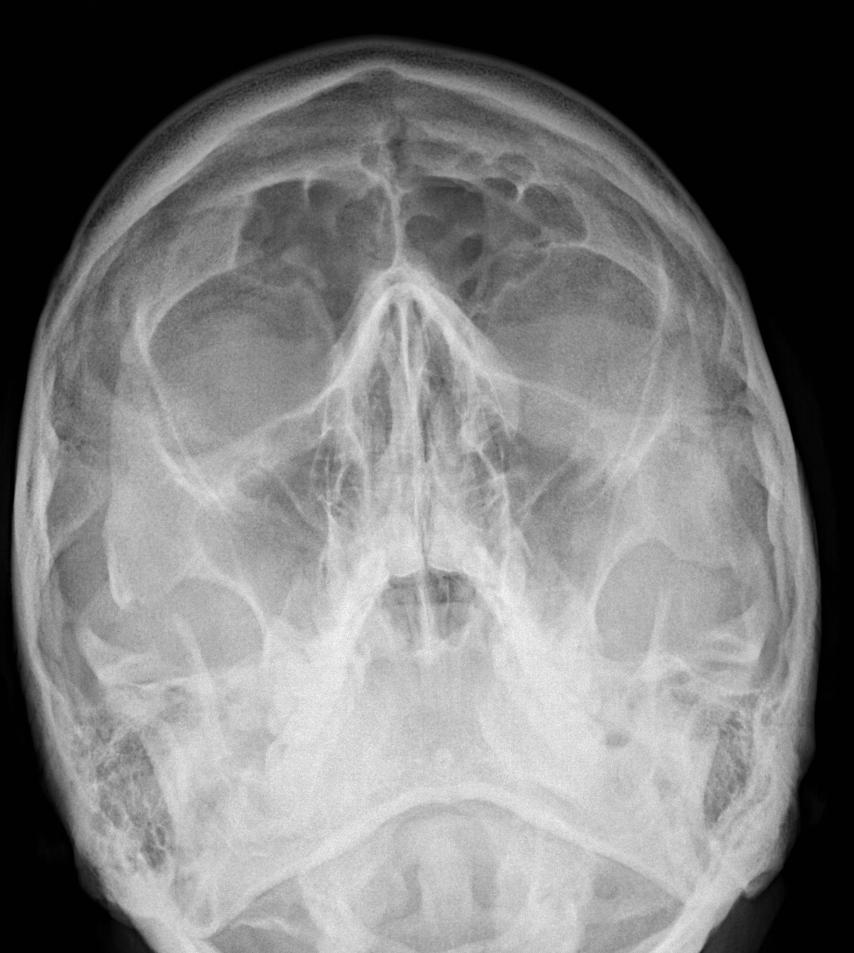

These complexes are referred to as the zygomaticomaxillary complex. The paired zygomas each have two attachments to the cranium, and two attachments to the maxilla, making up the orbital floors and lateral walls. The cause is usually a direct blow to the malar eminence of the cheek during assault.

The zygomatic arch usually fractures at its weakest point, 1.5 cm behind the zygomaticotemporal suture. Facial bruising, periorbital ecchymosis, soft tissue gas, swelling, trismus, altered mastication, diplopia, and ophthalmoplegia are other indirect features of the injury. In most cases, there is loss of sensation in the cheek and upper lip due to infraorbital nerve injury. On physical exam, the fracture appears as a loss of cheek projection with increased width of the face.

Its specific locations are the lateral orbital wall (at its superior junction with the zygomaticofrontal suture or its inferior junction with the zygomaticosphenoid suture at the sphenoid greater wing, separation of the maxilla and zygoma at the anterior maxilla (near the zygomaticomaxillary suture), the zygomatic arch, and the orbital floor near the infraorbital canal. The zygomaticomaxillary complex fracture, also known as a quadripod fracture, quadramalar fracture, and formerly referred to as a tripod fracture or trimalar fracture, has four components, three of which are directly related to connections between the zygoma and the face, and the fourth being the orbital floor. 2008 16(3):163–6.Right zygomaticomaxillary complex fracture with disruption of the lateral orbital wall, orbital floor, zygomatic arch and maxillary sinus. Economic analysis of open approach versus conventional methods of zygoma fracture repair. 187–8.Ĭzerwinski M, Ma S, Motakis D, Lee C. Surgery of the eyelid, orbit, and lacrimal system. 2012 23(3):859–62.Ĭornelius CP, Gellrich N, Hillerup S, Kusumoto K, Schuber W. Modified Gillies approach for zygomatic arch fracture reduction in the setting of bicoronal exposure. Swanson E, Vercler C, Yaremchuk M, Gordon C. Evidence based medicine: zygoma fractures. A textbook of advanced oral and maxillofacial surgery. Closed treatment of zygomatic arch fractureĪktop S, Gonul O, Satilmis T, Garip H, Goker K.The main complication of this procedure is facial asymmetry or contour deformity. An elevator is inserted through this incision, navigated to a location medial to the zygomatic arch, and used to reduce the zygomatic arch to its anatomic position. In the Gillies approach, a temporal incision is made anterior and superior to the root of the helix and carried through to a plane just deep to the superficial layer of the deep temporal fascia. Isolated zygomatic arch fractures yielding significant contour deformity or trismus are managed surgically (Ophthalmology 109:1207–1210, 2002). Isolated arch fractures are often the result of a direct lateral force to the arch that drives the fragments of the zygomatic arch medially (Surgery of the eyelid, orbit, and lacrimal system, Oxford, 212–217, 220–222, 1995). It serves as an attachment point for the masseter and plays a significant role in maintaining facial contour. The zygomatic arch is formed by the articulation of the temporal process of the zygoma and the zygomatic process of the temporal bone.


 0 kommentar(er)
